Micro-CT analysis can be used in various applications in dental research, such as enamel thickness, root canal morphology, root canal preparation, craniofacial bone structure, microscopic finite element modeling, dental tissue engineering, dental hard tissue mineral density and Implants and other aspects. It provides high-resolution images as well as qualitative and quantitative analysis of teeth, bones and implants.
Example 1: Adult teeth
Example 1: Adult teeth
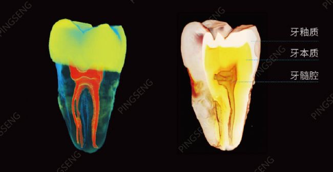
Figure 1 The Avatar software is a separate and accurate segmentation of the tooth enamel, dentin and pulp cavity, and provides accurate quantitative analysis and visualization.
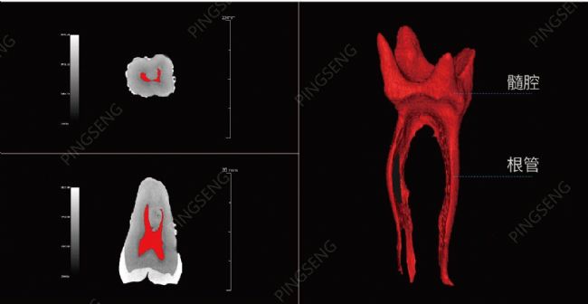
Figure 2 The segmented view and 3D rendering of the pulp cavity (the shape of the pulp cavity is roughly similar to the shape of the tooth, the medullary cavity of the crown is larger, called the pulp chamber, and the medullary cavity of the root is smaller, called the root canal)
The root canal is a kind of pore, and this low-density space in the middle of the tooth has a searchable direction for the study of endodontic disease. Micro-CT is particularly suitable for the three-dimensional quantitative evaluation of root canal fillings in the study of dental fillers.
The root canal is a kind of pore, and this low-density space in the middle of the tooth has a searchable direction for the study of endodontic disease. Micro-CT is particularly suitable for the three-dimensional quantitative evaluation of root canal fillings in the study of dental fillers.

Figure 3: Enamel as a high density material can be directly separated by a threshold. (The caries on the side of the test sample occur)
Enamel thickness has taxonomic and phylogenetic values ​​in human evolution. The effective and non-destructive technical properties of micro-CT are used to measure the enamel thickness of various archaeological specimens. In clinical studies, enamel thickness is considered to be important for the interpretation of the occlusion load protocol.

Figure 4: Avatar software can also generate continuous slices that accurately and reliably display the thickness and area of ​​enamel, dentin and pulp cavity. (a. adult true tooth photo, gray tone processing; b. horizontal slice display; c. vertical slice display)
Example 2: Rat mandible and caries
Rat or mouse mandible and caries are of great value in many research models of periodontal disease and other dental related fields. The micromanifold measurement of the mandible and teeth of the animal can further analyze the correlation between the characteristics of periodontal biotypes, and provide a theory for the evaluation of oral aesthetics, the choice of implant treatment, the judgment of treatment prognosis and the evaluation of curative effect. basis
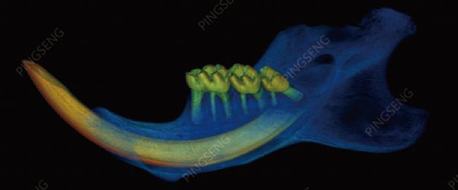
Figure 5: 3D rendering of rat mandible and caries
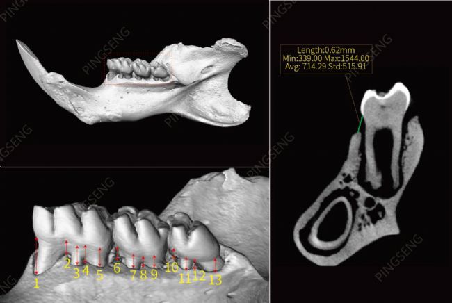
Figure 6: The maximum and minimum distance points from the alveolar process of each tooth to the gingival margin are selected and measured on a single slice.
Table 1: Distance of the alveolar to the gingival margin measured on a specific slice.

Table 1: Distance of the alveolar to the gingival margin measured on a specific slice.

Figure 7: a1, b1, c1, and d1 are the top cut planes of area a, b, c, and d, respectively, and a2, b2, c2, and d2 are the bottom cuts of several areas respectively; the rightmost figure shows The a, b, and c areas are all available on the surface, showing the size of the region of interest and the density of the trabecular bone in the region after the hand-drawn irregular ROI, standard elliptical ROI, and standard square ROI are displayed on the Avatar software. Mean, standard deviation, and maximum and minimum values. At the same time, other parameters of the trabecular bone of the region of interest can be quantitatively analyzed.
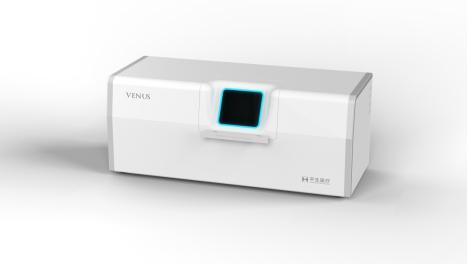
Experimental equipment: VENUS® Micro-CT
Chinese name: desktop high resolution micro CT
Model: VNC-100
Experimental equipment: VENUS® Micro-CT
Chinese name: desktop high resolution micro CT
Model: VNC-100
Imaging software: Avatar 1.3 (Pingsheng Medical Technology)
Teeth Whitening Machine,Desktop Model Tooth Light,Teeth Bleaching Machine ,Dental Teeth Whitening Machine
Foshan Ja Suo Medical Device Co., LTD , https://www.fjoralinstrument.com