Model NO.: RCS-500
Group: Female
Medical Device Regulatory Type: Type 2
Trademark: Carejoy
Transport Package: Carton
Specification: 56*46*31
Origin: Beijing, China
Model NO.: RCS-500
Group: Female
Medical Device Regulatory Type: Type 2
Trademark: Carejoy
Transport Package: Carton
Specification: 56*46*31
Origin: Beijing, China
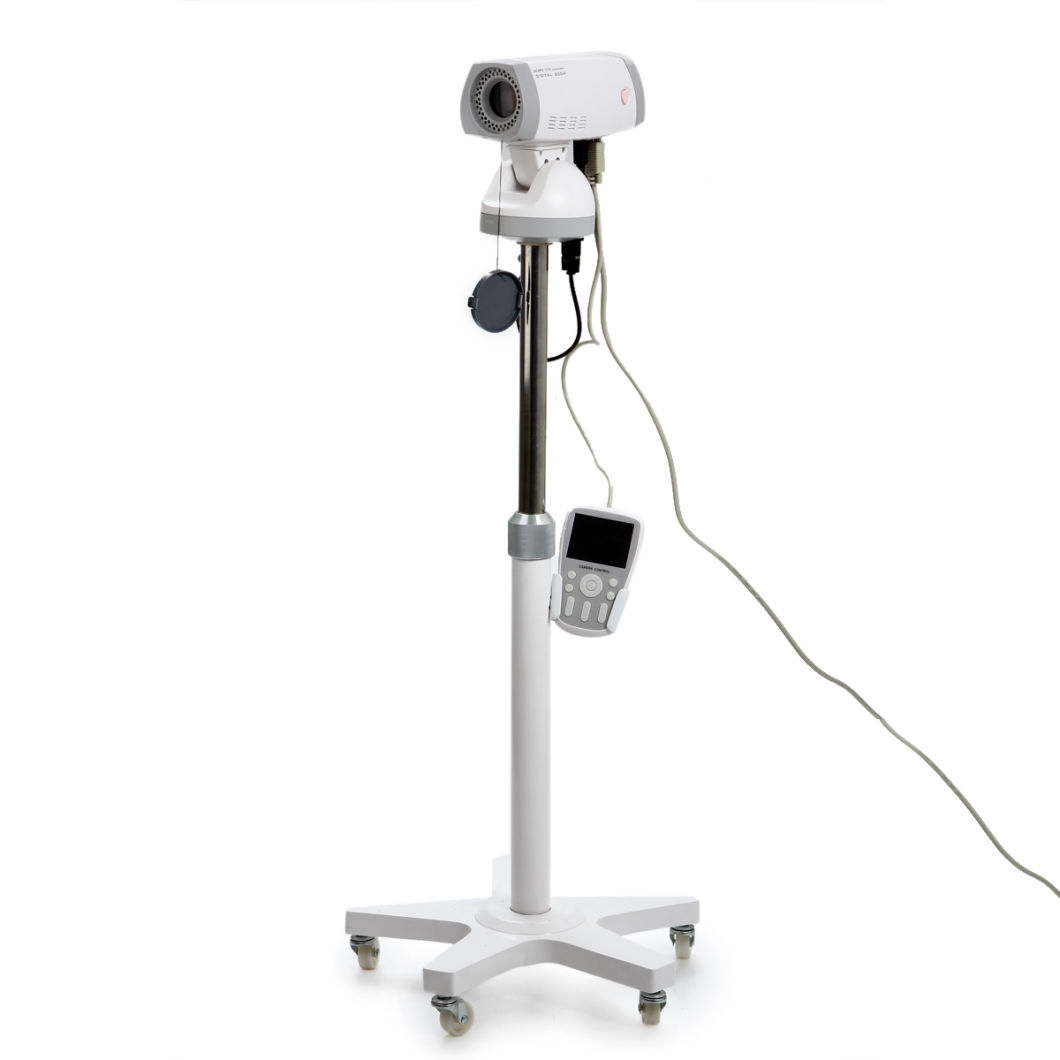
Product DescriptionRCS-500: SONY Camera 830,000 pixelsWork with Laptop & Desktop
Working Principle:Â
RCS-500 Digital Video Colposcope is applicable to detect pathological change in Vulva, vagina and the cervix, filtering cervix cancer by fluorescence indirectly, and images management.
Application:Â
For clinical detection of pathological changes in vulva, vagina and cervis, filtering cervis cancer by fluorescence indirectly and images management.
Feature of Performance:Â
The outlook is fashionable and convenient.
We adopt the color digital CCD camera of high resolution and clarity. (Sony)
The Colposcope is the sole dismountable light source, which has been got the patent.
Bulbs adopt supper cold LED light source whose diameter is 3 mm. And in one light source there are 30 bulbs.
There are freeze, filter, zoom in, zoom out, white balance button on the top of the camera for the speedy automatic function.
Professional green-filter technique
Image freeze
White balance
Auto focus
Timing of acetic acid and iodine reaction tests
Powerful image software and workstation system
Feature of Software:Â
Capacity of freezing and storing static images continuously and a maximum of 99 images available for reviewing simultaneously in scroll.
Capacity of cine loop and record continuously for different frequencies dynamic image in sampling.
Capacity of comparison of 4 pictures in the screen, multi-orientation and dynamic consultation, copy, freeze, and other function.
Supply hundreds of pathological reference image. Can help doctor compare the pathology and make the diagnoses.
Need not be inputted because of abundant diagnosis information
Capacity of internet report, and transmit the data through the internet to make an long-distance consultation and share of the information
Color image report in multi-formats and freely input information to make the user template
Tens of image enhanced functions to improve the checking rate of micro-focus
Sole Filter function to make sure to see the vein clearly with less loss of the light.
Easy operating software with icon, which can freeze and store 99 images consecutive and multi-orientation and dynamic consultation
Specification:
| CCD | 1/4 Â Super HAD CCD |
| Pixels | ≥830,000 pixel |
| Illumination (min): | ≤0.01Lux |
| Lens: | Sony 18 times focus |
| Resolution | ≥480+ lines |
| Light source | 30-point ringed LED, lili-white, super bright, cool, parallel, shadowless, 100, 000 hours longevity |
| Focus | 10mm ~400mm |
| Zoom magnification | Optical ≥54X, Eelectronical ≥12X |
| Collection model | Electronic collection |
| Depth of field | 5-200mm |
| Focalizing | Auto/Manual, quick |
| Signal-to-Noise | ≥52 db |
| Color filter | Green |
| Image collection | Y |
| Cineloop | Y |
| Output port of the image | -Video/PAL/NTSC |
| Green Filter | Digital filter based on CCD imaging |
| Eliminate polarization | Y |
| Field of vision | Φ=2.5-320mm |
| Software package | Y |
| Operating system | Windows 2000, XP, VISTA |
Standard configuration:
Image capture system
Image control system
Bracket
light source
Software
Plastic packing case
Optional configuration:
Foldable trolley
PC: desktop or latptop
 Product Description
RCS-500: SONY Camera 830,000 pixelsWork with Laptop & Desktop
Working Principle:Â
RCS-500 Digital Video Colposcope is applicable to detect pathological change in Vulva, vagina and the cervix, filtering cervix cancer by fluorescence indirectly, and images management.
Application:Â
For clinical detection of pathological changes in vulva, vagina and cervis, filtering cervis cancer by fluorescence indirectly and images management.
Feature of Performance:Â
The outlook is fashionable and convenient.
We adopt the color digital CCD camera of high resolution and clarity. (Sony)
The Colposcope is the sole dismountable light source, which has been got the patent.
Bulbs adopt supper cold LED light source whose diameter is 3 mm. And in one light source there are 30 bulbs.
There are freeze, filter, zoom in, zoom out, white balance button on the top of the camera for the speedy automatic function.
Professional green-filter technique
Image freeze
White balance
Auto focus
Timing of acetic acid and iodine reaction tests
Powerful image software and workstation system
Feature of Software:Â
Capacity of freezing and storing static images continuously and a maximum of 99 images available for reviewing simultaneously in scroll.
Capacity of cine loop and record continuously for different frequencies dynamic image in sampling.
Capacity of comparison of 4 pictures in the screen, multi-orientation and dynamic consultation, copy, freeze, and other function.
Supply hundreds of pathological reference image. Can help doctor compare the pathology and make the diagnoses.
Need not be inputted because of abundant diagnosis information
Capacity of internet report, and transmit the data through the internet to make an long-distance consultation and share of the information
Color image report in multi-formats and freely input information to make the user template
Tens of image enhanced functions to improve the checking rate of micro-focus
Sole Filter function to make sure to see the vein clearly with less loss of the light.
Easy operating software with icon, which can freeze and store 99 images consecutive and multi-orientation and dynamic consultation
Specification:
| CCD | 1/4 Â Super HAD CCD |
| Pixels | ≥830,000 pixel |
| Illumination (min): | ≤0.01Lux |
| Lens: | Sony 18 times focus |
| Resolution | ≥480+ lines |
| Light source | 30-point ringed LED, lili-white, super bright, cool, parallel, shadowless, 100, 000 hours longevity |
| Focus | 10mm ~400mm |
| Zoom magnification | Optical ≥54X, Eelectronical ≥12X |
| Collection model | Electronic collection |
| Depth of field | 5-200mm |
| Focalizing | Auto/Manual, quick |
| Signal-to-Noise | ≥52 db |
| Color filter | Green |
| Image collection | Y |
| Cineloop | Y |
| Output port of the image | -Video/PAL/NTSC |
| Green Filter | Digital filter based on CCD imaging |
| Eliminate polarization | Y |
| Field of vision | Φ=2.5-320mm |
| Software package | Y |
| Operating system | Windows 2000, XP, VISTA |
Standard configuration:
Image capture system
Image control system
Bracket
light source
Software
Plastic packing case
Optional configuration:
Foldable trolley
PC: desktop or latptop
Â
Trolley Ultrasound Scanner,Color Doppler Ultrasound,Doppler Ultrasound Diagnostic Instrument,Trolley Full Digital Ultrasound Scanner
Mianyang United Ultrasound Electronics Co., Ltd , https://www.ultrasonicequip.com