  LaVision BioTec Light-Sheet Microscopy Light Illumination Microscope Literature brief - VIPAR, a quantitative approach to 3D histopathology applied to lymphatic malformations background: In the process of drug development and surgical treatment of lymphedema and other lymphatic diseases, understanding of the mechanization of its mechanisms and mechanisms is particularly important. However, the lack of effective research and diagnostic tools has greatly limited this process. Moreover, in the study of true pathological changes in lymphatic vessels, standard two-dimensional tissue sectioning methods do not achieve the desired results. method: Volumetric Information Histopathology Analysis (VIPAR) for 3D reconstruction and data extraction is a method for analyzing tissue samples based on fluorescence microscopy, which is achieved by digital reconstruction and visualization of the microscopic image library. The VIPAR allows for semi-automatic segmentation of the vessel and subsequent unbiased extraction of characteristic vessel shapes and connection parameters. In this study, we used VIPAR to analyze healthy lymphoid and lymphangioma skin samples. result: Digital 3D reconstruction provides an intuitive, comprehensive representation of the lymphatic vessels and blood vessels in the tissue. The most striking feature found based on this technique is the disordered lymphatic drainage in the lymphedema skin and the proliferation of lymphangioma skin (4.36 times the increase in lymphatic vessel volume). Both abnormalities were found by linkage analysis based on extracted vessel morphology and structural data. Quantitative assessment of the acquired data revealed a significant reduction in lymphatic vessel length in lymphedema skin, a significant decrease in lymphatic flatness in lymphangioma skin, and a significant increase in blood vessel length in lymphangioma. All 3D imaging was performed by the LaVision light microscope with the software Imaris. Detailed introduction of LaVision light microscope: http://qd-china.com/products2.aspx?id=434 Example image of the result: VIPAR is a research and analysis method based on the imaging and visualization of light film, plus the basic steps of three 3D tissue reconstructions of segmentation, skeletonization and feature extraction. We use it for visualization, 3D reconstruction and quantitative analysis of microvessels in the skin. VIPAR analysis of vascular beds is based on semi-automated vessel segmentation to extract characteristic vascular parameters in a completely unbiased manner. The figure below shows the process of sample preparation, imaging and VIPAR method analysis. 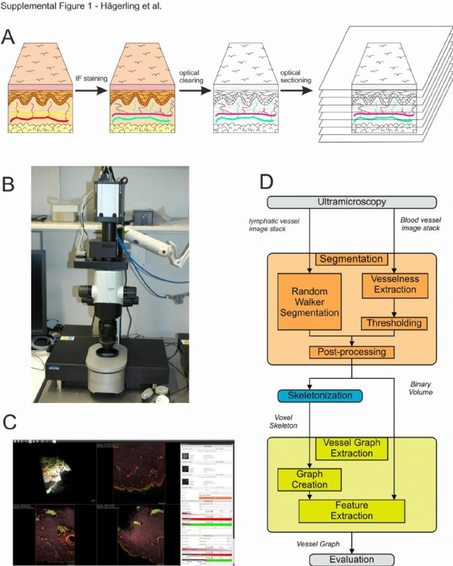 Results Figure 1. Three-dimensional visualization of the spatial arrangement of human dermal and lymphatic vessels using a light microscopy showed that visible lymphoids were visible in the papillary layer of the dermis (Fig. 1C, red arrow) and the reticular layer (Fig. 1C, green arrow). Cong. Both lymphatic plexus are connected by a bundle of vessels that are not continuously distributed, and these bundles of blood vessels are usually accompanied by lymphatic vessels (Fig. 1A). By observing the front of the nipple plexus through the skin, we immediately discovered the blind-end capillary lymphatic vessels (represented by asterisks, B, F, and H Figure 1) and the areas of dense lymphatic endothelial cells that express PROX1 (in B, F, and H is marked with a white arrow (Fig. 1)). Such dense structures (G and H in Figure 1) frequently appear near the lymphatic bifurcation and are additional evidence that they represent the hypothesis of lymphatic vessels. 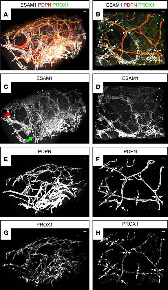 Results Figure 2 uses a light microscopy to analyze healthy and diseased skin samples and found lymphatic defects that are not recognized by conventional sectioning methods. We performed the same staining for healthy individuals (Figure 1 and Figure 2, D, G, J), lymphedema individuals (Figures 2, E, H, and K) and lymphangioma individuals (Figures 2, F, I, and L) The skin of the legs was subjected to VIPAR analysis to compare the effects of traditional histopathology and VIPAR. The 3D reconstruction process of VIPAR immediately showed lymphangiogenesis (compare Figures 2, D and G, Figure 2, E and H, Figure 2, F and I), consistent with histopathological analysis (Figure 2, AC).  Figure 3 The VIPAR method can detect differences in lymphatic features in healthy and pathological skin specimens. 3D reconstruction of PDPN positive lymphatic vessels showed significant differences between healthy skin and lymphedema and lymphangioma skin (Figures 3A-3C). Three-dimensional visualization of the lymphatic vessels showed that differences in lymphatic vessel density, volume, and morphology were preserved. The characteristics of irregularly arranged lymphatic vessels in lymphedema and the proliferation of lymphangioma are easily identifiable in the segmented lymphatic vessels (Fig. 3D-3I). To quantitatively assess changes in pathological samples, we used topological features extracted from the binary image generated by segmentation (Fig. 3J-3L) to calculate topological features, including the branch points of the lymphatic vessels and their connectivity (Fig. 3M-3O).  Figure 4: Automatic segmentation of blood vessels in the experimental and illuminating groups in digital three-dimensional reconstruction. Evaluation of vascular system based on ESAM1 immunostaining (Fig. 5, AC): semi-automatic segmentation of blood vessels by calculating features on multiple Gaussian scales, then manual global thresholding to obtain binary images, followed by post-processing, and removal of small The segmentation error. The segmented vascular structure is represented as a binary body (Fig. 5, DF). In the subsequent skeletalization process, the central axis of each vessel was calculated by stereoscopic thinning (Fig. 5, GI).  in conclusion: In summary, VIPAR has proven to be suitable for the analysis of two vasculature systems in human skin samples. It can detect significant defects and quantitatively assess the pathological state of lymphedema and lymphangioma with unbiased data extraction and morphological parameter calculations. Quantum Quantum Scientific Instruments Trading (Beijing) Co., Ltd., as the exclusive partner of LaVision BioTec GmbH in China, we will continue to provide the latest product application cases and cutting-edge information for the majority of scientific research users. Quantum Quantum Science Instruments Trading (Beijing) Co., Ltd. Website: http:// mailbox: Beijing Office Room 0306-0308, 3/F, Air China Building, 36 Xiaoyun Road, Chaoyang District, Beijing 100027 Phone: 010-8512 0277/78/79/80 Fax: 010-8512 0276 Shanghai Office Room 1405, Shanghai International Group Building, 511 Weihai Road, Jing'an District, Shanghai 200041 Phone: 021-5228 0980 Fax: 021-5228 2156 Guangzhou Office Room 1712, R&F Yingfeng Building, No. 2 Huaqiang Road, Zhujiang New Town, Tianhe District, Guangzhou 510623 Phone: 020-8920 2739 Fax: 020-8920 2750 |
China Pigment Suppliers
Here you can find the related products in Pigment, we are professional manufacturer of Pigment. We focused on international export product development, production and sales. We have improved quality control processes of Pigment to ensure each export qualified product.
If you want to know more about the products in Pigment, please click the Product details to view parameters, models, pictures, prices and Other information about Pigment.
Whatever you are a group or individual, we will do our best to provide you with accurate and comprehensive message about Pigment!
Lycopene, Amaranth, β-Carotene, Beta carotene, Carmine, Food Colorant
Xi'an Gawen Biotechnology Co., Ltd , https://www.ahualyn-bio.com