(1) What is the "chemotaxis movement"?
"Chemotaxis" is a directed movement of cells under chemical stimulation (chemokines), including "Chemotactic migration" and "Chemotactic invasion", which is an immune response. An important component of physiological phenomena such as cell differentiation, tumor metastasis, wound healing and blood vessel formation. Studying the "chemotaxis movement" of cells is critical to further understanding the biological processes described above.
(2) Defects of traditional research methods
Traditional "chemotaxis" studies were performed using the "Boyden chamber", which was observed at the end of chemotaxis. Although the “Boyden chamber†can study the response of cells to chemokines, it has obvious defects: it cannot obtain dynamic images of migration and invasion of cells, and requires a large number of cells (50,000 to 100,000 cells/well). The most important is Only the “end point method†can be used for observation, and the dynamic process of “chemotaxis†of cells varies greatly with different cell types and chemokines. The “end point method†cannot distinguish different dynamic processes at all.
(C) Essen 's "ClearView Chemotactic Microplate"
Essen BioScience has developed innovative technologies for dynamic, image-based, high-throughput 96- well “chemotaxis†research. The technology utilizes the "ClearView 96-well chemotaxis microplate" containing an optically transparent membrane as a consumable, and can perform fully automatic dynamic imaging of the entire process of "chemotaxis" in the "IncuCyte Live Cell Dynamic Imaging and Analysis System". During this process, the cells are maintained in a physiological environment in a standard incubator.

Figure 1. “ClearView 96-well chemotaxis microplate†dedicated to “chemotaxisâ€. The plate consists of three parts: the upper layer is a transparent cover, the middle layer is a 96-well plate for planting cells, and the lower layer is a transparent reagent tank. In the 96-well plate of the middle layer, there is an optically transparent film in each of the holes, and a plurality of precise small holes having a diameter of 8 μm are printed by laser. During the experiment, cells were seeded into the membrane in the wells of the middle plate, and Chemoattractant was injected into the reagent tank of the lower layer. Then, the "IncuCyte Live Cell Dynamic Imaging and Analysis System" was placed to dynamically record the movement of cells through the pores under the action of chemokines.
(4) How to quantify the “chemotaxis movement�
Full-hole imaging of the upper and lower surfaces of the film in the "ClearView Chemotaxis Microplate" at user-defined intervals was performed using an automated algorithm to quantify the cell area of ​​the upper and lower surfaces (Figure 2). For adherent cells, an increase in the cell area of ​​the lower surface of the membrane can be calculated; for suspended cells, a reduction in the cell area of ​​the upper surface of the membrane can be calculated, at which time the cells fall through the pores on the membrane below the membrane.
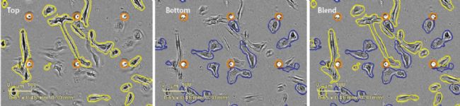
Figure 2. Unlabeled analysis of “chemotaxisâ€. The seeding density of HT-1080 fibrosarcoma cells was 1,000 cells/well. The "chemokine" in the lower reagent tank is 10% FBS. The left side is the upper surface of the 36-hour membrane (the cells are hooked out in yellow); the middle is the 36-hour lower surface of the membrane (cells are drawn in blue), and the right side is a composite view of the upper and lower surfaces. Orange red indicates a small hole in the membrane.
(5) Advantages of IncuCyte's “chemotaxis†solution
The IncuCyte “chemotaxis†solution has many distinct advantages over the traditional “Boyden chamber transwells†and “microfluidic devicesâ€, such as:
(1) Visualize the dynamic cell migration/invasion process, output dynamic images, and observe cell morphology;
(2) Long-time automated analysis in the incubator, retaining cell growth status, saving manpower, and outputting a dynamic curve of migration/invasion;
(3) Highly repeatable 96-well dynamic experiment suitable for screening;
(4) can be used as adherent cells, but also as suspension cells;
(5) The cells can be cultured separately or co-cultured;
(6) Compatible with label - free analysis and fluorescent label analysis;
(7) Low cell dosage , suitable for rare cells, expensive cells or primary cell populations. The ClearView plate requires 1,000-5,000 cells per well per well, while the "Boyden chamber" method requires a minimum of 50,000 cells per well per well;
(8) The density of the pores is smaller, the cells need to move on the surface of the membrane, and are surface-mediated migration/invasion processes, which can express the matrix properties of the membrane surface and deeper phenotypic characteristics;
(9) The concentration gradient of chemokines on both sides of the membrane can be maintained for a longer period of time. The ClearView method maintains the concentration gradient for more than 72 h, and a significant concentration gradient drop can be observed after 4 h of the "Boyden chamber" method.
(6) Characteristics of IncuCyte's “chemotaxis†solution
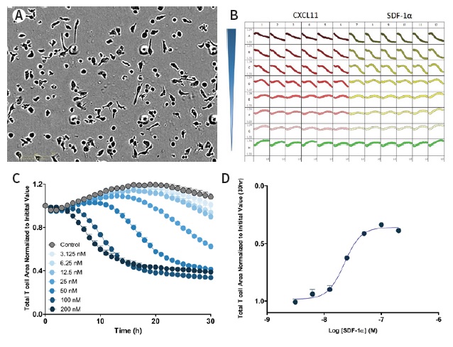
Figure 3. Image and data analysis of real-time 96-well "chemotaxis". Panel A: High-resolution phase contrast image of T cells on the IncuCyte ClearView 96-well plate. Panel B: 96-well plate curve of T cells against different concentrations of chemokines CXCL11 and SDF-1α. Panel C: Dose response curve of T cells to different concentrations of chemokine SDF-1α, the ordinate is the cell area of ​​the plate. The greater the visible concentration, the faster the curve drops, indicating that the upper cells pass through the small holes quickly. Panel D: plot the dose response curve at the data point of 30 h to obtain a plot of the logarithmic value of the upper T cell area versus the SDF-1 alpha concentration.

Figure 4. The 96- well “chemotaxis†curve shows excellent inter-plate repeatability. The Z's value of the four microplates was 0.5-0.7 in three days, and the corresponding concentration response curve to the chemokine SDF-1α showed reproducibility of response to SDF-1α (EC50 of 19-33 nM).

Figure 5. Detection of "surface matrix" mediated cell migration. The ClearView plate has a very low pore density, and neutrophils must migrate on the surface of the membrane in order to move toward the chemokine. Left: The membrane of the ClearView plate was not coated, and the cells did not migrate. The chemokines were IL-8 and fMLP. Right: The surface of the membrane is coated with Matrigel, and the cells have obvious chemotactic movement. This data shows that the interaction of integrin and/or cell surface receptors with the matrix is ​​key to the neutrophil chemotaxis in this model. In contrast, when neutrophil chemotaxis was performed in "Boyden chamber transwells", no coating was required (data not shown).
(7) Classification of “chemotactic movementâ€
"Chemotaxis" can be divided into (1) "chemotaxis migration" across the surface of the substrate; (2) "chemotaxis invasion" through 3D biomatrix and (3) transthoracic "chemotaxis migration" of leukocytes.
7.1 "chemotaxis migration" through the surface of the substrate
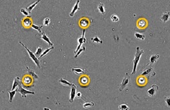
Figure 6. "Chemotactic migration" across the surface of the substrate
feature:
(1) Dynamically monitoring and quantifying the targeted cell migration of cells through plastic plates or biological substrates;
(2) Suitable for a wide range of immune, tumor and vascular cell types;
(3) The effect of different treatment methods on the full-time of “chemotaxis migration†can be studied.
7.2 “Chemical invasion†through 3D biomatrix
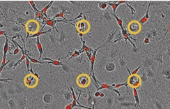
Figure 7. "Chemotactic invasion" through 3D biomatrix
feature:
(1) Dynamically imaging and quantifying the "chemotaxis invasion" of cells through 3D biomatrix, such as "tumor invasion" ;
(2) can assess the potential of metastasis and define the impact of treatment methods on the invasive phenotype ;
(3) Different biological processes of “chemotaxis migration†and “chemotaxis invasion†can be explored on the same microplate.
7.3 "chemotaxis migration" of leukocytes through the endothelium
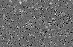
Figure 8. "Chemotactic migration" of leukocytes through the endothelium
feature:
(1) monitoring and quantifying the dynamic migration of leukocytes through the endothelial monolayer;
(2) Visualize the integrity of the endothelial monolayer and leukocyte exudation ;
(3) Exploring the biological effects of inhibitors and neutralizing antibodies.
(8) Summary
Compared to the traditional "Boyden chamber transwells" and "microfluidic devices", the IncuCyte "chemotaxis" solution has obvious advantages for various "chemotaxis" experiments.
(9) Automated live cell dynamic analysis
At present, most of the cell detection methods still belong to the traditional end point method - only the final result is given. This method can not only dynamically monitor and analyze the whole process of cell experiments, but also obtain biased results. In order to be able to observe live cells throughout the process, Essen Corporation of the United States has developed the IncuCyte S3 "Viable Cell Dynamic Imaging and Analysis System" built into the incubator, inside the incubator, and simultaneously for 6 cell culture dishes/bottles/microplates (6 -384 wells) for long-term dynamic imaging, recording real-time changes in cell morphology details, and transforming into a variety of cell function-related time-quantitative curves through powerful image analysis software.
IncuCyte S3 is a cell-level scientific and drug screening solution for immunosuppression, nerve growth and regression, cancer cell metastasis, cell migration, stem cell differentiation, apoptosis, cell necrosis, cell proliferation, wound repair, etc. Large-scale screening and research on long-term life science issues.
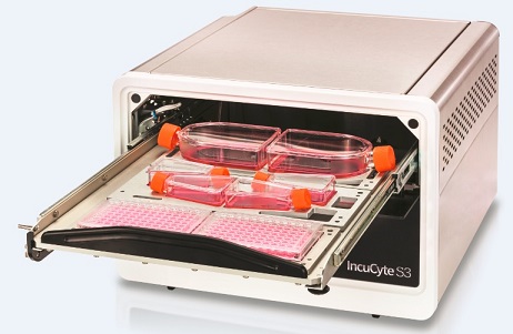
Figure 9. IncuCyte S3 Live Cell Dynamic Imaging and Analysis System
(10) About "Diaoao Biotechnology"
“ Diao Biotech †(Shanghai, Hong Kong, Taiwan) was established in 1994. For more than 20 years, it has been adhering to the world-class instrument manufacturers. The experts with rich R&D experience are technical support and the professional maintenance team serves. Backed by a focus on providing state-of-the-art hardware and software equipment and technical services for life sciences, pharmaceutical R&D and disease treatment.
The exclusive distributors of " Diao Biotech " are:
IncuCyte, Beacon, Chipcytometry, Gyrolab, iQue Screener PLUS, Avatar, PlexBio, Certus, HighRes, etc.
Telephone (transfer to the marketing department)
Email:
Website:
Scan the QR code to add the WeChat public account " tekontech " :

Vitamins are a type of trace organic substances that humans and animals must obtain from food in order to maintain normal physiological functions. They play an important role in human growth, metabolism and development. Vitamins do not participate in the formation of human cells in the body, nor do they provide energy for the human body. Vitamins are a class of organic compounds necessary for maintaining good health.
Our company provides group A vitamins, group B vitamins, B12, etc.

Vitamin Tablet,Food Grade Vitamin,Vitamins For Energy,Water Soluble Vitamins
XI AN RHINE BIOLOGICAL TECHNOLOGY CO.,LTD , https://www.rhinebioteches.com